


Cutting-edge imaging sheds new light on cells that break down bone
Bone may seem as if it’s a hard, lifeless structure, but now the cells living within have been imaged in unprecedented detail, thanks to an innovative imaging method developed at the Garvan Institute of Medical Research. Credit: Garvan Institute Cutting-edge imaging sheds new light on cells that break down bone Imaging technology developed at Garvan […]

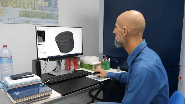
Bone may seem as if it’s a hard, lifeless structure, but now the cells living within have been imaged in unprecedented detail, thanks to an innovative imaging method developed at the Garvan Institute of Medical Research.

Credit: Garvan Institute
Cutting-edge imaging sheds new light on cells that break down bone
Imaging technology developed at Garvan shows that bone-resorbing osteoclasts gather in distinct pockets, leading to new insights for osteoporosis and cancer treatment.
Bone may seem as if it’s a hard, lifeless structure, but now the cells living within have been imaged in unprecedented detail, thanks to an innovative imaging method developed at the Garvan Institute of Medical Research.
The new method lets researchers study cells inside the bones of mice, to visualise not just isolated sections, but the entire length of a bone. With a new level of visual detail, the researchers discovered that osteoclasts, cells that break down bone tissue, are more active in some parts of the bone compared to others. This knowledge could be used to develop new treatments for osteoporosis, and for dormant cancer cells, which can stay hidden in bone for years until they are reactivated by osteoclasts.
“Our method has given us an unprecedented window into how cells go about breaking down bone, giving us a new way to investigate osteoporosis and cancer relapse in bone,” says Professor Tri Phan, Head of the Intravital Microscopy Lab and Gene Expression (IMAGE) Lab, immunologist at St Vincent’s Hospital Sydney, Co-Director of the Precision Immunology Program at Garvan and senior author of the paper, published in Nature Protocols.
“We can finally image processes inside bone that we thought were happening, but which were until now beyond the limits of conventional microscopy techniques. We are only beginning to understand the implications of this exciting technology.”
Giving disease-causing cells no place to hide
Osteoclasts are crucial to the normal maintenance and repair processes of bone, but when they are overly active, they can cause excessive breakdown, known as osteoporosis.
“The inside of living bone is a ‘dark space’ that is difficult to study, because of its hard, mineralised structure,” says co-first author Dr Nayan Deger Bhattacharyya, post-doctoral researcher in the IMAGE Lab. “In order to understand diseases such as osteoporosis and cancer recurrence, we’ve needed to develop the technology to look inside bone tissue.”
The new technique developed at Garvan’s ACRF INCITe Centre can be used to image other dynamic cellular processes until now hidden in bone.
“Our new imaging method is minimally invasive and lets us map out localised populations of cells along the length of an entire bone in our mouse models, instead of just in small sections,” says co-first author Wunna Kyaw, PhD student in the IMAGE Lab.
The researchers tracked down distinct pockets of bone resorption activity as the cells ‘morph’ between actively resorbing osteoclasts and an intermediate cell state called osteomorphs, in real time.
“We suspect these osteomorphs are dangerous as they can accumulate while osteoporosis treatment is administered but can rapidly reform activated osteoclasts to supercharge bone breakdown as soon as treatment is stopped. This would explain an observation in the clinic, that many osteoporosis patients taking the medication denosumab, which blocks osteoclasts from resorbing bone, experience rebound vertebral fractures after they stop using the drug. We will use our imaging method to study how this withdrawal effect could be prevented,” says co-author Professor Peter Croucher, Head of the Bone Biology Lab at Garvan.
The researchers say their method could also be used to investigate cancer cells that can migrate to bone during cancer treatment and lie dormant there for years, only to be reactivated by osteoclasts breaking down the bone tissue that surrounds them.
“Being able to see cells and molecules interact in the bone – and one day target them – could be a critical new tool for diseases relating to bone,” says Professor Phan.
–ENDS–
This research was supported by the Australian National Health and Medical Research Council (NHMRC) grants ID1155678 and ID2009010, the Mrs Janice Gibson and Ernest Heine Family Foundation, Mr Peter Duncan AM and the late Mrs Val Duncan, Kedje Foundation, Cancer Institute New South Wales (CINSW) and the Australian Cancer Research Foundation (ACRF).
Professor Tri Phan is a Conjoint Professor at St Vincent’s Clinical School, Faculty of Medicine and Health, UNSW Sydney. Professor Peter Croucher is a Conjoint Professor at St Vincent’s Clinical School, Faculty of Medicine and Health, UNSW Sydney.
Journal
Nature Protocols
DOI
10.1038/s41596-023-00894-9
Method of Research
Observational study
Subject of Research
Animals
Article Title
Minimally invasive longitudinal intravital imaging of cellular dynamics in intact long bone
Article Publication Date
19-Oct-2023
What's Your Reaction?