Revolutionary MIT Technique Labels Protein in Millions of Densely Packed Cells Within Organ-Scale Tissues
A groundbreaking advance in the realm of biological research has emerged from the laboratories at the Massachusetts Institute of Technology (MIT). Recent innovations have enabled scientists to label proteins across millions of individual cells within intact three-dimensional tissues, using sophisticated techniques that promise a new era of molecular biology exploration. The technology, showcased in a […]


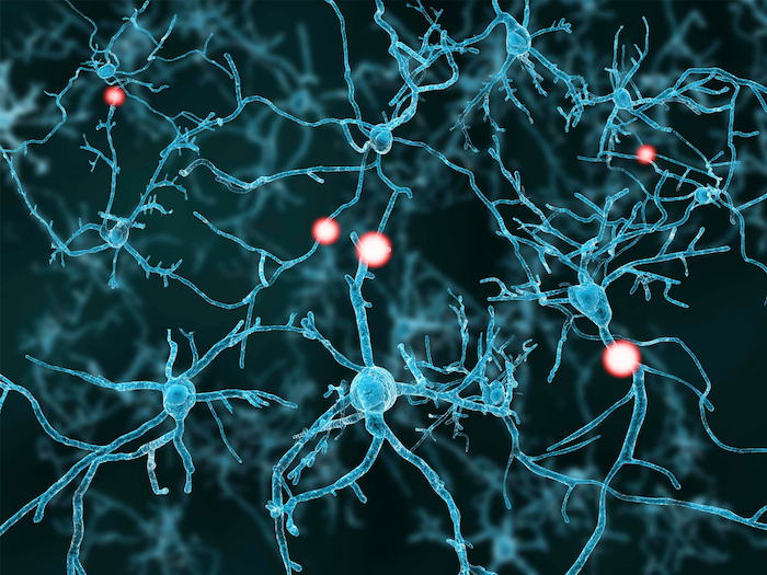
A groundbreaking advance in the realm of biological research has emerged from the laboratories at the Massachusetts Institute of Technology (MIT). Recent innovations have enabled scientists to label proteins across millions of individual cells within intact three-dimensional tissues, using sophisticated techniques that promise a new era of molecular biology exploration. The technology, showcased in a study published in Nature Biotechnology, significantly enhances the ability to investigate protein expression across entire organs, including rodent brains and other complex tissues, all within a dramatically shortened time frame of just a single day.
Traditionally, researchers face substantial limitations when attempting to analyze protein expression within intricate three-dimensional biological structures. Current methodologies often necessitate the dissociation of tissue into single cells or sectioning them into thin slices for analysis. This process not only risks losing vital context but also tends to overlook the holistic interactions between cells within their native environments. Consequently, many cellular functions, especially in relation to disease or therapeutic responses, remain obscured due to the inadequacies of existing technologies.
The development of this new labeling technology aims to address these shortcomings head-on. By employing an innovative technique known as “CuRVE,” the research team at MIT has made significant strides toward achieving uniform labeling throughout large and densely packed tissues. This approach not only allows for comprehensive protein identification across entire organs but does so with remarkable speed and accuracy. The key to CuRVE lies in its ability to control the antibody binding kinetics while simultaneously accelerating antibody penetration into the tissue. This dual approach brings forth a fundamental shift in how researchers can study complex biological systems, turning the daunting task of cellular analysis into a feasible endeavor.
In their pioneering study, the researchers have successfully demonstrated that their enhanced protein labeling technology can be implemented without the extensive optimization typically required for different types of tissues. This represents a significant leap in efficiency for the field of molecular biology. They experimented with over sixty different antibodies, achieving successful labeling results across a variety of specimens, including whole mouse brains, embryos, and other organs like the lungs and hearts. Given that these techniques can uniformly process large tissue volumes within single-day durations, the implications for both basic research and clinical applications are staggering.
The practicality of this technology is further underscored by the collaborative efforts behind its development. Innovators from MIT’s Picower Institute for Learning and Memory have been at the forefront of this pioneering research, particularly Kwanghun Chung, who has long been involved in creating methodologies that visualize biological samples in unprecedented detail. The concept of CuRVE draws inspiration from previous techniques such as CLARITY and SWITCH, both of which focused on tissue transparency and targeted protein binding, respectively.
While the innovations underlying CuRVE are remarkable, the real genius lies in its applications. By integrating strategies that separate antibody binding and permeation, researchers can actively modulate the speed at which antibodies attach to their target proteins while promoting their movement through dense tissue. This is akin to adjusting the marinade absorption process of a thick steak — ensuring that every part of the tissue uniformly takes up the labeling agents. This analogy anchors an important takeaway about the technology: it doesn’t simply label proteins but does so in a manner that accurately reflects the true biological state of the tissue being studied.
This systematic capability allows researchers to confidently explore new avenues of investigation. For instance, the relentless pursuit of understanding the brain’s complexities poses vast opportunities for discovering neuronal interactions and activities that were previously inaccessible. By providing insights from the contextual entirety of cellular interactions, this technology may enhance our understanding of neurological disorders, paving the way for innovative treatment strategies that leverage precise knowledge of protein dynamics.
Moreover, the discoveries facilitated by CuRVE and its eFLASH implementation emphasize its superior performance compared to conventional genetic tagging approaches. While genetic methods offer a means to track protein expression through fluorescent markers, discrepancies often exist between the transcriptional processes and actual protein levels. The MIT team observed significant variations when comparing these methods, highlighting the robustness of antibody labeling for accurately identifying protein presence in a spatial context that genetic methods lack.
As this investigation progresses, the scientific community’s discourse on protein labeling methods will likely expand significantly. Researchers will now possess a powerful tool that can bridge the previously existing gaps between single-cell analysis and whole-organ investigations. The interdisciplinary nature of this research underscores its relevance across numerous domains, from neuroscience to cancer research, enhancing the understanding of vital biological interactions and clinical phenomena.
The funding that supported this pivotal study underscores its significance, with various esteemed entities contributing resources to foster such groundbreaking research. The collaborative efforts among scholars and institutions reflect a broader commitment to advancing the life sciences, ensuring that substantial discoveries can translate into tangible benefits for public health.
In conclusion, the potential ramifications of this technology extend far beyond the confines of academic research. With the ability to examine proteins across various animal models rapidly and comprehensively, this method represents a paradigm shift in biological investigation, enabling scientists to address fundamental questions about life at cellular and molecular levels. This research opens up exciting possibilities for future explorations in biology, promising new insights that can ultimately drive innovation in medicine and healthcare.
Subject of Research: Animals
Article Title: Uniform volumetric single-cell processing for organ-scale molecular phenotyping
News Publication Date: 24-Jan-2025
Web References: Nature Biotechnology
References: Nature Biotechnology
Image Credits: Credit: Chung Lab/MIT Picower Institute
Keywords
Life sciences, Biochemistry, Proteins, Neuroscience, Molecular biology, Protein expression, Biomedical engineering, Imaging, Brain research.
Tags: advancements in biological researchcellular interactions in diseaseCuRVE technology for proteinshigh-throughput protein analysisintact tissue protein labelingMIT protein labeling techniquemolecular biology innovationsNature Biotechnology studynovel biomedical techniquesorgan-scale tissue researchprotein expression investigationthree-dimensional tissue analysis
What's Your Reaction?


