


Innovative Method for Detecting “Aged” Cells Opens New Frontiers in Ageing Research
Researchers at Tokyo Metropolitan University have pioneered a groundbreaking label-free technique that allows for the precise differentiation of “aged” or senescent human cells from their younger counterparts through the application of alternating electric fields. Conventional methods for identifying senescent cells typically depend on biochemical labeling, such as fluorescent tags that bind to specific markers unique […]


Researchers at Tokyo Metropolitan University have pioneered a groundbreaking label-free technique that allows for the precise differentiation of “aged” or senescent human cells from their younger counterparts through the application of alternating electric fields. Conventional methods for identifying senescent cells typically depend on biochemical labeling, such as fluorescent tags that bind to specific markers unique to aged cells. While these established approaches have advanced understanding, they also come with significant limitations: the labeling process is labor-intensive, time-consuming, and, crucially, the labeling itself can alter the very cellular properties researchers aim to investigate. The novel electric field-based method circumvents these drawbacks, offering a minimally invasive, faster, and more reliable strategy to study cell aging.
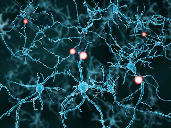
At the heart of this scientific breakthrough lies the recognition that ageing is fundamentally a cellular process. As organisms age, their tissues accumulate cells that have ceased to divide and perform their original functions—known as senescent cells. These cells don’t merely become inert; they actively secrete pro-inflammatory compounds that contribute to pathologies associated with ageing, including arterial stiffening, neurodegenerative diseases such as Alzheimer’s, and metabolic disorders like type 2 diabetes. Understanding how these senescent cells behave and influence tissue function is essential to devising interventions to prevent or treat age-related diseases. However, the challenges in accurately identifying these cells without altering their biology have significantly hampered research progress.
The innovative technique, spearheaded by Assistant Professor Ippei Yagi and his team, employs what is known as frequency-modulated dielectrophoresis (FM-DEP), involving the exposure of cells to an alternating electric field. Unlike static electric fields, the alternating nature produces dynamic cellular responses. When placed in such a field, cells experience a slight redistribution of electric charges, leading to the phenomenon where one side of the cell becomes more positively charged relative to the other end. Importantly, when the electric field is spatially non-uniform, this induced polarization causes cells to move—a behavior termed dielectrophoresis. In alternating electric fields, such cells oscillate between electrodes, and their motion is frequency-dependent. By systematically varying the frequency, researchers observe a characteristic threshold called the ‘cutoff frequency’ at which cell movement markedly changes.
.adsslot_PAri1GgUbl{ width:728px !important; height:90px !important; }
@media (max-width:1199px) { .adsslot_PAri1GgUbl{ width:468px !important; height:60px !important; } }
@media (max-width:767px) { .adsslot_PAri1GgUbl{ width:320px !important; height:50px !important; } }
ADVERTISEMENT
This cutoff frequency serves as a biophysical fingerprint, reflecting intrinsic electrical and structural properties of the cell. The research team applied FM-DEP primarily to human dermal fibroblasts—connective tissue cells critical for maintaining skin integrity. Experimentation revealed a pronounced and reproducible difference in cutoff frequency profiles between young and senescent fibroblasts. This distinction arises from biochemical alterations, chiefly in the lipid composition of cellular membranes, that accompany ageing. Membrane lipids, which contribute to cellular electrical properties, undergo modification during senescence, thereby affecting how cells respond to external electric stimuli. This coupling between membrane biochemistry and electrophysical properties enables FM-DEP to distinguish cell age effectively.
Beyond its scientific novelty, FM-DEP shines in practical terms. The method is both rapid and straightforward, eliminating the need for staining or molecular tagging. This attribute not only preserves cell viability and native biological states but also simplifies experimental protocols, potentially accelerating research cycles. Importantly, the label-free aspect makes FM-DEP highly adaptable for living cell studies in real-time and may facilitate high-throughput screening platforms.
The implications of this technique extend far beyond mere cellular identification. By enabling precise discrimination of senescent cells, FM-DEP paves the way for improved understanding of cellular senescence’s role in tissue degeneration and systemic ageing. This knowledge is vital for advancing regenerative medicine strategies, where the rejuvenation of aged tissues or removal of senescent cells could restore function in damaged organs. Moreover, the method shows promise for drug screening applications to identify compounds that specifically target or modulate senescent cells without deleterious side effects on normal cells.
Intriguingly, the team envisions broadening FM-DEP’s scope to encompass various cell types beyond dermal fibroblasts. Such versatility would offer an unprecedented tool to study ageing in diverse tissues, potentially unraveling distinct electrophysiological signatures among senescent cells depending on their origin. It might also enable detection of early senescent changes before phenotypic markers become evident, thus enhancing diagnostics.
Technically, the FM-DEP setup involves a microfluidic device with electrodes generating spatially non-uniform alternating electric fields across a suspension of cells. As the frequency sweeps, electrodes induce a time-dependent dielectrophoretic force on cells, measured through their migration velocities and oscillatory behavior. The cutoff frequency is quantitatively determined by analyzing the frequency at which motility patterns shift, providing a reproducible indicator of cellular state. This quantitative nature facilitates objective classification, potentially deployable in automated cell sorting systems.
The scientific community has long grappled with the challenge of minimally invasive detection of cell senescence without disrupting physiology. FM-DEP’s reliance on biophysical characteristics rather than biochemical markers represents a paradigm shift. It leverages fundamental physics to elucidate complex biological phenomena, exemplifying the power of interdisciplinary innovation between engineering and life sciences.
This research aligns with a growing trend toward label-free cellular analysis, including techniques such as impedance spectroscopy and optical tweezing, yet FM-DEP distinguishes itself by targeting the dynamic interplay of frequency-dependent dielectric properties. Its success underscores the importance of exploring electrical properties of cells as a rich source of information about cell health, phenotype, and function.
Funded by JSPS KAKENHI under grant numbers JP23K28453 and JP23KK0260, this work stands as a testament to the fruitful collaboration between physicists, biologists, and engineers at Tokyo Metropolitan University. With publication in the IEEE Sensors Journal scheduled for June 11, 2025, the research community awaits further developments and applications of FM-DEP with great anticipation.
As the population ages globally, technologies like FM-DEP could revolutionize how we monitor and combat ageing-related diseases, ultimately extending healthy lifespan. By facilitating rapid, reliable, and non-invasive detection of senescence, this method brings us closer to a future where personalised interventions in tissue ageing become routine clinical practice.
Subject of Research: Cellular senescence and label-free identification of senescent human dermal fibroblasts using electric fields
Article Title: Label-free Detection of Senescence-like State in Human Dermal Fibroblasts via Frequency-Modulated Dielectrophoresis
News Publication Date: 11 June 2025
Web References: http://dx.doi.org/10.1109/JSEN.2025.3576789
Image Credits: Tokyo Metropolitan University
Keywords: Cellular senescence, Dielectrophoresis, Fibroblasts, Biophysics, Biomedical engineering, Cell biology, Electrical properties, Electrodes, Regenerative medicine
Tags: Aging Researchcellular aging and healthconventional senescence identification limitationselectric field technology in biologyimplications of aged cells in diseasesinnovative methods in cellular biologyinterventions for age-related pathologieslabel-free cell differentiationminimally invasive research techniquespro-inflammatory compounds in agingsenescent cell detection methodsTokyo Metropolitan University research
What's Your Reaction?