Connecting the dots to shape growth forces
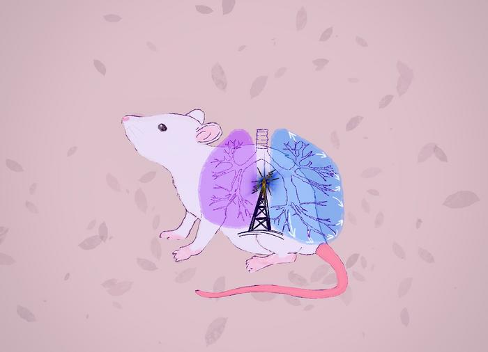
Kyoto, Japan — Branching patterns are prevalent in our natural environment and the human body, such as in the lungs and kidneys. For example, specific genes that express growth factor proteins are known to influence the development of the lungs’ complex branches. Still, until now the mechanics behind this phenomenon have remained a mystery. Credit: […]

Kyoto, Japan — Branching patterns are prevalent in our natural environment and the human body, such as in the lungs and kidneys. For example, specific genes that express growth factor proteins are known to influence the development of the lungs’ complex branches. Still, until now the mechanics behind this phenomenon have remained a mystery.

Credit: KyotoU Tobiyama / Tsuyoshi Hirashima
Kyoto, Japan — Branching patterns are prevalent in our natural environment and the human body, such as in the lungs and kidneys. For example, specific genes that express growth factor proteins are known to influence the development of the lungs’ complex branches. Still, until now the mechanics behind this phenomenon have remained a mystery.
Kyoto University researchers have unveiled a regulatory system linking signal, force, and shape in mouse lung structure development. The team recognized that the signal protein ERK plays an active role in causing growing lung tissue to curve.
“ERK signals the cell tissue to stretch outward to smoothen its curve,” says Tsuyoshi Hirashima, formerly of KyotoU’s Graduate School of Biostudies and now at the National University of Singapore’s Mechanobiology Institute.
As if choreographed, a mix of chemical signals triggers the cellular mechanics of the lungs of a mouse embryo, resulting in the development of intricate branching patterns.
Mechanobiology has gained increasing attention in recent years, focusing on cell- and tissue-generated forces, intracellular signaling, and their combined interactions with geometric factors that influence morphogenesis.
“ERK’s surprisingly precise signaling response to lung tissue curvature was enlightening. It suggests an elegantly more nuanced developmental orchestration than previously thought,” reflects Hirashima.
Utilizing advanced microscopic imaging techniques, Hirashima’s team observed how ERK behaves in developing lungs in real time by combining a fluorescent biosensor — for quantifying the ERK activity in living cells — with two-photon microscopy, which captures tissue cell and molecular activities in 3D.
Results showed that ERK mediates curvature sensing and force generation in epithelial cells, causing a negative feedback loop and a repetitive branching pattern.
“We are particularly interested in exploring how disruptions in this signal-force-shape system might contribute to physiological abnormalities or diseases,” says Hirashima.
These ideas may apply to the developmental processes of other organs and the formation of mouse lungs, a realization that calls for further exploration of fundamental principles.
“Ultimately, our findings offer a deeper understanding of the novel principles of biological regulatory systems, with promising applications in regenerative medicine and organoid research,” concludes Hirashima.
###
The paper “ERK-mediated Curvature Feedback Regulates Branching Morphogenesis in Lung Epithelial Tissue” appeared on 15 January 2024 in Current Biology, with doi: 10.1016/j.cub.2023.12.049
About Kyoto University
Kyoto University is one of Japan and Asia’s premier research institutions, founded in 1897 and responsible for producing numerous Nobel laureates and winners of other prestigious international prizes. A broad curriculum across the arts and sciences at undergraduate and graduate levels complements several research centers, facilities, and offices around Japan and the world. For more information, please see: http://www.kyoto-u.ac.jp/en
Journal
Current Biology
DOI
10.1016/j.cub.2023.12.049
Method of Research
Experimental study
Subject of Research
Cells
Article Title
ERK-mediated Curvature Feedback Regulates Branching Morphogenesis in Lung Epithelial Tissue
Article Publication Date
15-Jan-2024
COI Statement
The authors declare no competing interests.
What's Your Reaction?


